Adult acquired flatfoot deformityor posterior tibial tendon(tibialis posterior) dysfunction
How does this deformity present?
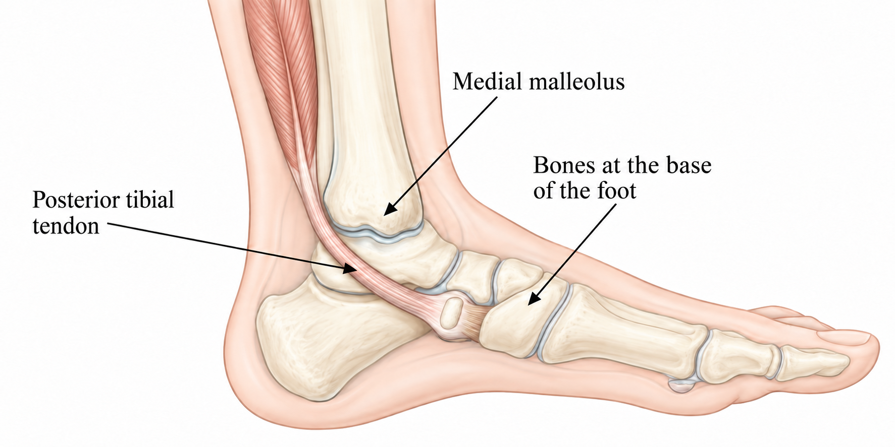
The posterior tibial tendon (tibialis posterior) (see illustration) helps support the arch of the foot and prevents excessive pronation, or outward deviation of the foot. It runs behind the inner ankle (medial malleolus) and attaches to the bones at the base of the foot. Sometimes this tendon, together with the ligaments supporting the arch, becomes excessively stretched and inflamed. This may cause progressive adult acquired flatfoot deformity. Correction requires a multidisciplinary team approach. Common names include posterior tibial tendon dysfunction, posterior tibial tendon insufficiency and adult acquired flatfoot deformity (AAFD).
Common causes
Flatfoot — most people who develop this deformity already have flat feet. Excessive or continuous loading causes the arch to flatten further, with pain and swelling on the inside of the ankle. Inadequate support from footwear may contribute.
Trauma or injury — the condition may sometimes be associated with a fracture, sprain or direct tendon injury.
Age — the risk of posterior tibial tendon dysfunction increases with age. Research indicates that it is most common in middle-aged women.
The following sequence of events is thought to cause AAFD:
A person with an existing flatfoot deformity places greater load on the tibialis posterior tendon, the main tendon supporting the arch. The strength, elasticity and endurance of muscles, tendons and ligaments diminish over a lifetime. In some people, the tendon gradually becomes overloaded, stretched, inflamed or torn. This is usually not sudden; it develops over months. As the tendon stretches, the arch ligaments also stretch and tear. Under body weight, the arch bones move out of position: the heel rotates outwards and the forefoot moves laterally, flattening the arch. The deformity may progress until the foot deviates outwards from the ankle.
Classification
Posterior tibial tendon dysfunction is characterised by worsening symptoms and deformity. It is classified into four stages.
Stage I:
- stretched tendon;
- pain around the outer ankle, especially when walking;
- swelling along the tendon;
- the patient can rise onto the toes of one leg;
- treatment uses insoles and physiotherapy.
Stage II:
- partial tendon tear;
- increasing pain and swelling;
- increased flattening of the foot;
- the patient cannot rise onto the toes of one leg;
- treatment is similar to Stage I, with tendon reconstruction if required.
Stage III:
- the heel is deviated outwards;
- treated with insoles or orthoses;
- surgical correction may be required — arthrodesis of the hindfoot joints or an osteotomy.
Stage IV:
- ankle deformity;
- ankle surgery may be required.
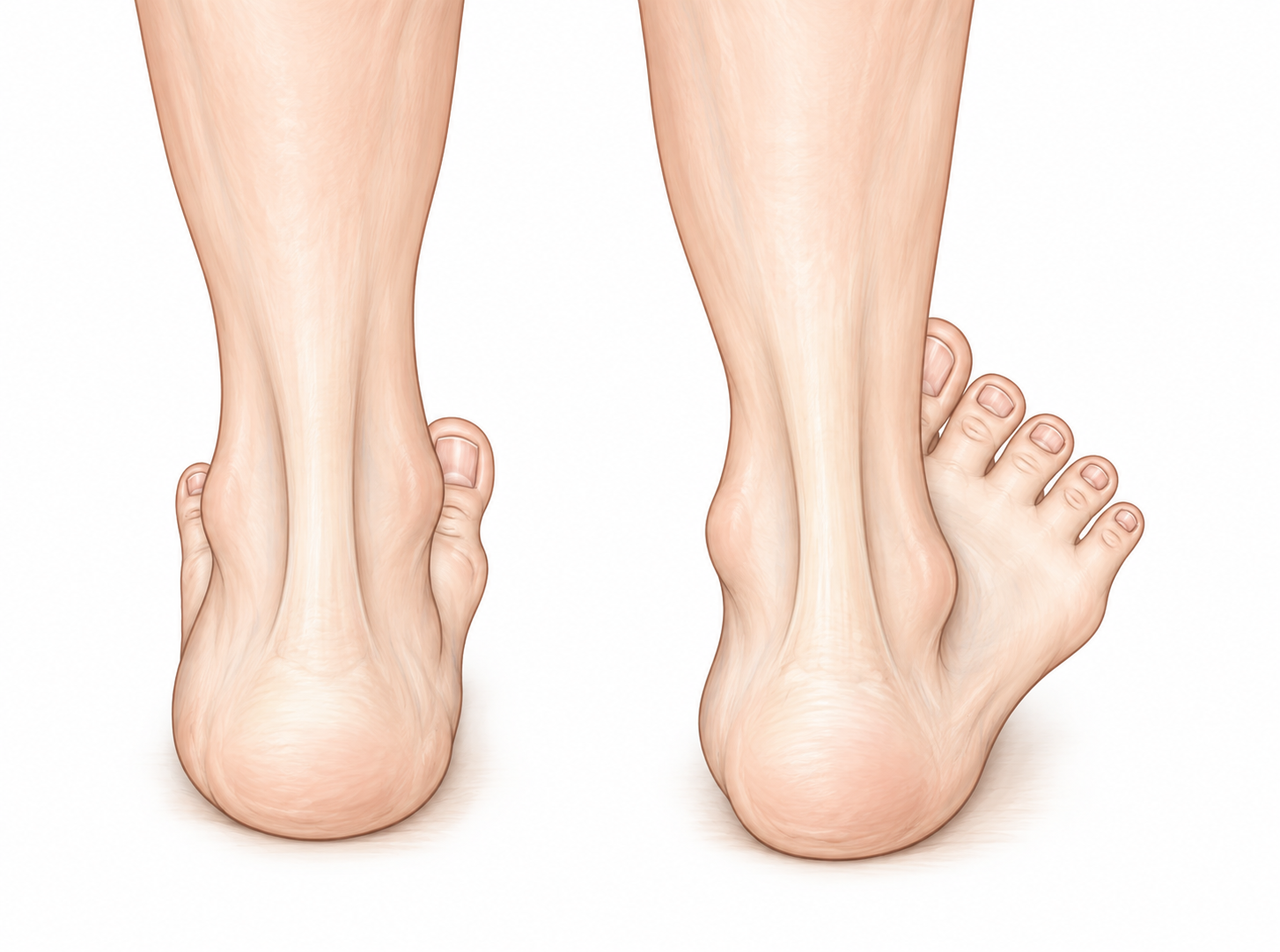
Compare the deformed right foot with the healthy left foot. The longitudinal or medial arch is flatter and the heel deviates outwards (valgus). More toes are visible on the right because the foot has moved outwards (abduction).
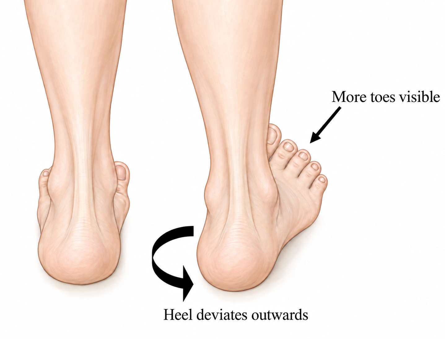
(see illustration) Together with inability or difficulty rising onto the toes, these signs indicate posterior tibial tendon dysfunction.
Common symptoms
Pain and/or swelling on the inside of the ankle and along the arch, following the course of the posterior tibial tendon. Pain may be burning, sharp or stabbing; tingling may occur from irritation of a nerve in the tarsal tunnel. Over time, pain and swelling may also develop on the outside of the ankle because the lower leg bone impinges on the heel bone. Pain may occur when moving the foot inwards or outwards and when stretching the foot.
Difficulty walking — inability to walk long distances or persistent pain when walking short distances. Pain may worsen at the end of the day.
Foot deformity develops as the tendon stretches. The tendon and ligaments weaken, the arch flattens and the shape of the foot changes.
Inability to walk on tiptoe is a very important diagnostic sign. The movement may cause pain.
Treatment
AAFD is best treated early. A precise and comprehensive assessment by your doctor will determine the most suitable treatment.
Conservative (non-surgical) treatment:
Activity should be reduced and the RICE regimen followed:
R — rest — rest as often as possible. Avoid activities that aggravate the condition, including exercise, sport and long walks.
I — ice — ice the painful area. Place a towel between the ice and the foot to protect the skin. Apply cold packs every two hours for 15–20 minutes.
C — compression — compression with an elastic bandage to reduce symptoms, pain and discomfort.
E — elevation — keep the leg elevated to reduce swelling.
Your doctor may prescribe pain medication — non-steroidal anti-inflammatory drugs, provided you have no allergy or asthma.
Immobilisation of the affected leg. Two options are available: a below-knee plaster splint for 4–8 weeks or a removable walking boot.
Appropriate footwear is important. Firm, lace-up, flat footwear such as trainers or shoes is recommended. It supports the foot and keeps orthotic insoles in the correct position.
Orthoses — insoles, functional orthoses, ankle supports and ankle-foot orthoses — are usually fitted individually to provide maximum functional stability. They improve tendon mechanics and reduce stretching, pain and inflammation.
Physiotherapy. Exercises and physiotherapy help improve tendon endurance and mobility and stretch the Achilles tendon, reducing pain.
In Stage I, inflammation and pain can usually be treated without surgery. Long-term use of insoles is needed to reduce recurrence or worsening symptoms. This is essential to prevent progression to the next stage.
In Stage II, the tendon is substantially stretched and the heel deviates outwards. Complete ankle immobilisation in a plaster splint or special boot may reduce symptoms and halt progression in a small percentage of patients. Long-term immobilisation may help control the deformity and pain without surgery.
In Stages III and IV, non-surgical treatment cannot stop progression but may be an alternative for patients who do not want surgery or cannot undergo it for health reasons.
Surgical treatment:
Surgery should be considered when ALL non-surgical methods have been used. Its aims are to relieve pain, stop progression and improve mobility. Specialists differ on the best method. Common procedures include surgical debridement of the tendon, tendon transfer, osteotomy and arthrodesis.
Surgery is rarely needed in Stage I. Early Stage II is treated with tendon surgery followed by heel-bone surgery.
If the tendon is substantially stretched or torn, a tendon replacement may be required. It can be replaced with another tendon, such as the flexor digitorum longus, or with part of the anterior tibial tendon in a Cobb procedure. This is performed through one or more incisions on the inside of the ankle.
The heel bone may be repositioned by calcaneal osteotomy. Through a lateral incision, the bone is moved into the correct position beneath the lower leg and fixed with a screw.
Additional procedures may sometimes be required to balance the foot, such as Achilles tendon lengthening or fusion of the transverse tarsal joint.
Arthrodesis of one or more joints may be required to correct Stage III or IV deformity.
Your surgeon may postpone the operation until you stop smoking. Smoking is also discouraged after surgery and while the bones are healing. Research shows that smoking delays bone healing.
Important advice after tendon and bone surgery (Stage II)
You will remain in hospital for 2–4 days. Treatment is individual, but every patient will have the leg immobilised in a plaster splint after surgery.
Your foot may be placed in an inverted position immediately after surgery. The plaster splint will be changed every two weeks to move the foot gradually into a neutral position.
Postoperative appointments — regular reviews with your treating doctor are required. The dressing is removed, the wound is examined and a new plaster splint is applied.
Until the foot is neutral, you must not bear weight on it and must use crutches while supporting yourself on the healthy leg. A physiotherapist will assess you in hospital and teach you how to use crutches correctly. Once the foot is neutral, you will be able to bear full weight when walking.
Prolonged physiotherapy is required after surgery. You may also need an insole.
Returning to work depends on your circumstances and occupation. If your work is sedentary and you can keep the operated foot elevated, you may return from the fourth postoperative week. Full recovery may take up to 12 months.
Driving — if the left foot was operated on and you drive an automatic car, you may drive from the fourth week. You must be able to perform an emergency stop. Tell your insurer about the operation and confirm that your policy remains valid.
Sport — walking and light exercise may be resumed, but strenuous sporting activity is not recommended after surgery.
Possible complications of surgical treatment
Modern foot surgery has a successful outcome in 90% of cases, but, as with any surgical treatment, there is a known risk of complications.
Infection develops in a small percentage of patients. Infected bone or a screw may need to be removed. Minor infections are more common and usually resolve after a short course of antibiotics.
Bleeding is common after surgery; you may notice blood on the dressing. Elevating the foot can reduce it.
Numbness and tingling result from damage to small nerves. Sensory changes are usually temporary but may be permanent.
Non-union — bones sometimes fail to unite. Smoking substantially increases the risk of non-union and other major complications. It is essential to stop smoking before surgery and avoid smoking until all bones have united.
Scars remain after any operation and may sometimes cause pain and irritation.
Prominent screw — a troublesome screw may occasionally need to be removed.
Blood clots — deep vein thrombosis (DVT) and pulmonary embolism (PE) are rare. Tell your doctor if you have had DVT or PE, or if your family has a history of clotting disorders.
Persistent pain and deformity — surgery may be unsuccessful in up to 15% of cases and further surgery may be required.
Recurrence of the deformity and symptoms is rare; repeat surgery can be performed if necessary.
CONTACT YOUR DOCTOR IF YOU DEVELOP SEVERE PAIN, MARKED SWELLING, EXCESSIVE NUMBNESS OR TINGLING.